THE SURGEON DECIDES.
RLES AI MAKES SURE NOTHING IS MISSED.
AI-Assisted Clinical Decision Support
for Cataract & Refractive Surgery
ANALYZE. DETECT. PLAN. EXPLAIN.
In clinical testing since January 2026 · Figures as of —.
Field observations from an ongoing clinical pilot — not a controlled trial.
SEE WHAT A SECOND ANALYTICAL LAYER FINDS.
Pilot surgeons shape the rule set: binding rules in the engine today came from questions asked by surgeons in this programme.
YOUR AI ASSISTANT
AT YOUR SIDE.
One mobile interface on your phone. No forms, no app-store install, no learning curve — between consultations or on your way out of the OR, RLES AI is with you.
Diagnostic reports, images, values or voice notes — in the RLES AI Mobile Interface.
Clinical parameters, corneal risk, surgical limits and device-specific data.
Potential contraindications and safety concerns — identified before planning.
An explainable, signature-ready surgical planning report.
The final clinical decision always belongs to the surgeon.
No paperwork. Just send and go.
No installations. No learning curve.
Expert-level analysis within minutes.
Two channels, one engine: the same rules, the same safety floors and the same reports, whichever way you reach them.
BEFORE PLANNING THE SURGERY,
RLES AI TRIES TO FIND A REASON NOT TO.
Every plan — no exceptions — passes through binding safety floors before it reaches your signature. Designed to catch the one case before it ever reaches the operating room.
NO RISK FLAG IS HIDDEN. EVERY WARNING CARRIES ITS CLINICAL RULE, CALCULATION AND REASONING.
SEE THE RISK
BEFORE THE LASER FIRES!
Fifteen clinical modules across four clinical territories — from the first ectasia screen to the cataract surgery decades later — plus three platform-wide features. Open only what you need; every capability has its own page.
One laser plan requires about 300 separate items to be asked, examined, read and calculated — a cataract plan about 210, for both eyes. Percent tissue altered, the single strongest marker of ectasia risk in eyes with normal topography, is not displayed by any device: it has to be calculated. See what one plan actually requires, with the evidence →
Laser Surgery Planning
Procedure-specific pre-operative planning for SMILE, LASIK, iLASIK, Q-LASIK, PRK and T-PRK — signature-ready in minutes.
Keratoconus & Ectasia Screening
Multi-index ectasia screening with correlation-aware analysis — runs even when device software says “Normal”.
Precision WFG / TG Planning
Plans by the source of astigmatism — corneal, lenticular or mixed — not just the manifest cylinder.
Enhancement Retreatment Planning
Stability checks and a cumulative tissue budget choose the safest second procedure for each eye.
Presbyopia Planning
Candidate screening and expectation management for every blended-vision strategy your devices can deliver.
Complication Prevention & Analysis
Detects the preconditions of free flap, buttonhole and ectasia at planning — and analyses adverse events without blame.
Keratoconus Treatment Planning
From consensus progression assessment to the right protocol — CXL ladder, CAIRS and brand-specific ring segments.
Contact-Lens Warpage Detection
Nine indicators catch lens-deformed corneas before an unstable map can mislead a plan.
Dry Eye Syndrome Analysis
Full DEWS II structured assessment — plus a silent tear-film screen on every topography you send.
Refractive Passport & IOL Support
A lifetime document for the patient today — and the right IOL inputs decades later.
Glaucoma Screening — NEW
Thirteen rules watch where anterior-segment surgery meets glaucoma — narrow angle, pigment, the post-refractive IOP trap — and close with a referral summary, never a diagnosis.
Phakic ICL Referral
When laser says no, EVO/EVO+ candidacy opens automatically — with the ACD measurement trap corrected.
Cataract & RLE Planning — NEW
The cornea's true power cross-validated through four tiers — never an IOL power, never a brand.
THE RIGHT LENS BEGINS WITH
THE TRUE POWER OF THE CORNEA.
Most refractive surprises after cataract surgery start with one wrong assumption: that a single keratometry number tells the truth. RLES AI replaces that assumption with cross-validation — and keeps learning from your own outcomes.
TRUE CORNEAL POWER ENGINE
Four independent measurement tiers, agreement bands and a confidence score on every case — disagreement is the finding, not noise.
CANDIDACY & RISK SCREENING
IOL candidacy at class level, posterior-surface astigmatism for the toric decision, and a full surgical risk profile.
FORMULAS & LENS DATA
Formula priority for virgin, post-refractive, short, long and keratoconic eyes — with an IOLCon-verified constant catalogue.
CALIBRATED TO YOUR HANDS
A personal C-constant and personal SIA built from your own outcomes — statistically gated, never guessed.
The boundary is absolute: RLES AI never calculates IOL power and never recommends a brand or model. It verifies the inputs, screens the candidacy and calibrates the process — the decision and the calculation remain yours.
Outcome Registry & Personal Nomogram
One-line follow-ups become your personal nomogram — proposals only when your own statistics can defend them.
XAI — Explainable AI
Every recommendation carries its rule, formula and literature — audit any number down to its source.
Voice Notes & Voice Report — NEW
Send voice notes, confirm every value, listen to voice reports — audio never leaves RLES AI's own server.
Patient Information Report
Your final decision, explained to the patient in plain language — never a recommendation.
Multi-Language Support — NEW
Work in your own language — analyses, reports and patient documents in sixteen languages.
EVERY DEVICE. EVERY SETTING.
ONE INTELLIGENT NOMOGRAM.
Register your clinic's devices once. From then on, every analysis applies the published nomograms and specifications of your platform — values are never substituted from another device.
EXCIMER LASERS
Published specifications and treatment-zone geometry, per platform.
MICROKERATOMES & FEMTO
Manufacturer nomograms · manufacturer tables · programmed femtosecond flaps.
DIAGNOSTIC DEVICES
Printed outputs are read and processed with device-specific reading cards. An off-catalogue model is no obstacle: values are read from its printout, and capability is never guessed.
EVERY WARNING HAS A RULE.
EVERY RULE HAS A SOURCE.
Every recommendation carries the rule it came from, the calculation behind it and the literature that supports it. Any number can be audited back to its source — and RLES AI never rules that surgery “cannot be done”; the decision always rests with the surgeon.
A DEVELOPMENT OF
MCD TECHNOLOGY INVESTMENTS INC.
RLES AI® is a development and investment of MCD Technology Investments Inc. — every rule is built in accordance with academic publications and the medical literature, and designed around the patient experience and clinical practice of experienced cataract and refractive surgeons. It is verified against published device nomograms and current literature, and delivered to physicians as clinical decision support. For professional use; the final clinical decision always rests with the surgeon.
RLES AI commercial launch: November 2026 — doctors and clinics wishing to join the pilot are being registered. This page is a preview of RLES AI®, intended for professional users only.




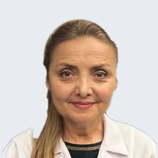

RLES AI's clinical rules are continuously challenged, refined and validated with practising ophthalmic surgeons.
THE FINAL DECISION
WILL ALWAYS BE YOURS.
RLES AI helps you see more before you make it.
APPLY TO USE RLES AI
The clinical pilot is open and free until the end of November 2026 — no licence, no commitment, no limit on places. Apply to use RLES AI with your own cases, or simply request information. Every application is reviewed personally; approved surgeons are registered on the RLES AI line the same day.